Normal mouse primary skeletal muscle cell culture
2020-10-29 14:15:55
PriCells - normal mouse primary skeletal muscle cell culture
First, experimental reagents
1. Medium: PriCells Medium + 10% FBS + 1% P/S + PriCells Supplement
2. Cryopreservation solution: PriCells Medium + 20% FBS + 10% DMSO
3. Washing solution: 1 × PBS (pH 7.4 ) + 1% P/S
4. Staining solution: 0.4% Trypan Blue
5. Digestive juice: PriCells Isolation of Primary Cell Kit
6, detection reagent: mouse anti-human and rat desmin primary antibody, myosin monoclonal antibody
Second, the experimental equipment
1. Petri dish
2, culture bottle
3, direct cut and eye scissors
4, ophthalmology and hemostats
5, beaker
6, 15ml centrifuge tube
Third, the experimental process
Take the muscles in the plate, Hank's wash 3 times
↓
Eliminate fat and connective tissue
↓
Muscle specimen cut about 0.1cm3
↓
Move to the centrifuge tube and wash Hank's 3 times
↓
Allow to stand for 1 min, discard the supernatant and floating tissue
↓
0.25% trypsin, digested in a 37-degree water bath for 20 minutes, shaking or blowing once every 5 minutes
↓
The growth medium is terminated, repeatedly blown, and sieved 100, 200, 400 mesh in sequence.
↓
Centrifugation, 1000rmp, 10min, discard the supernatant
↓
Resuspend, count cells, inoculate density at 5 × 10 5 /ml
↓
The suspension was added to the culture flask without PPL coating, and the fibroblasts were removed by differential adhesion, 37 degrees, 1 hour.
↓
Transfer the bag to the flask, culture in the growth medium, culture at 37 ° C, 5% CO 2
↓
4d change, change every day
Fourth, experimental operation
1. The newborn mouse is sacrificed by pulling the cervical vertebrae, and the legs are sterilized by ethanol. In the ultra-clean workbench, the muscles are taken in the plate, Hank's is washed 3 times, the fat and connective tissue are removed, and the muscle specimen is cut into small pieces of 0.1 cm3;
2. Move the cut muscles to the centrifuge tube, wash Hank's 3 times, let stand for 1 min, discard the supernatant and floating tissue.
3. Add 0.25% trypsin to the above centrifuge tube, digest for 20 minutes in a 37-degree water bath, shake or blow the centrifuge tube once every 5 minutes, and then add the growth medium to terminate the digestion;
4, after repeated blowing, 100,200,400 mesh in sequence, the filtrate was collected, centrifuged at 1000r / min for 10min;
5. Discard the night, resuspend the cells in growth medium, add the suspension to the culture flask without PPL, remove the fibroblasts by differential adherence, incubate at 37 °C for 1 h, transfer the coating to the flask, and grow and culture. Base culture, 4d change of liquid, and change the liquid once a day.
V. Cell identification
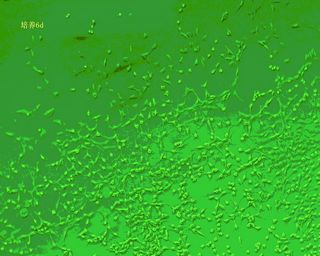
1. Microscopic identification: The primary cells that have just been isolated are relatively small, spherical, and have strong refractive index. After 12h, the cells began to adhere to the wall. After 72h, the cells adhered completely, and the cells gradually expanded into a fusiform shape. As the culture time prolonged, the cells proliferated and migrated and gradually arranged in one direction. When the cells are fused more than 80%, the myotubes begin to form.
2. Immunocytochemical staining 1: Desmin is contained in the cytoplasm of muscle satellite cells, and the satellite cells can be immunostained and identified by mouse anti-human and mouse desmin primary antibodies.
3. Immunocytochemical staining 2: Detection by skeletal muscle-specific myosin monoclonal antibody. The skeletal muscle cell coverslips were routinely treated and subjected to immunochemical staining of myosin cells, and PBS was used as a negative control instead of primary antibody. The results were judged: brown staining of cytoplasm appeared positive staining.
Six, matters needing attention
1. In the process of trypsin digestion, it can also be carried out by a two-step digestion method of 15 minutes. According to the age of the individual tissues taken, the adult adult mice have a good two-step digestion effect, and the young mice have little difference in one-step digestion and two-step digestion.
2, not too much subculture algebra, skeletal muscle cell passability is limited, easy to die.
3. During skeletal muscle cell culture, the morphology will change greatly.
4, muscle tissue should remove the surface of the membrane and fat tissue as much as possible.
Seven, PriCells cell picture
Fried Carrot Flakes,Vacuum Fried Carrot,Dried Carrot,Bulk Carrot
Huaiyang County Wanyuan Garlic Foods Processing Industries Co.,Ltd , https://www.wanyuangarlicfood.com